
Telemedical diagnosis of retinopathy of prematurity. Scott KE, Kim DY, Wang L, Kane SA, Coki O, Starren J et al. AMIA Annu Symp Proc, American Medical Informatics Association 2010 2010: 111–115. Diagnostic performance of a telemedicine system for ophthalmology: advantages in accuracy and speed compared to standard care. Am J Ophthalmol 2011 152 6: 1053–1058 e1.Ĭhiang MF, Wang L, Kim D, Scott K, Richter G, Kane S et al. Accuracy and reliability of telemedicine for diagnosis of cytomegalovirus retinitis. Ophthalmology 2012 119 (3): 617–624.Īusayakhun S, Skalet AH, Jirawison C, Ausayakhun S, Keenan JD, Khouri C et al. Quality of nonmydriatic digital fundus photography obtained by nurse practitioners in the emergency department: the FOTO-ED study.

Lamirel C, Bruce BB, Wright DW, Delaney KP, Newman NJ, Biousse V.

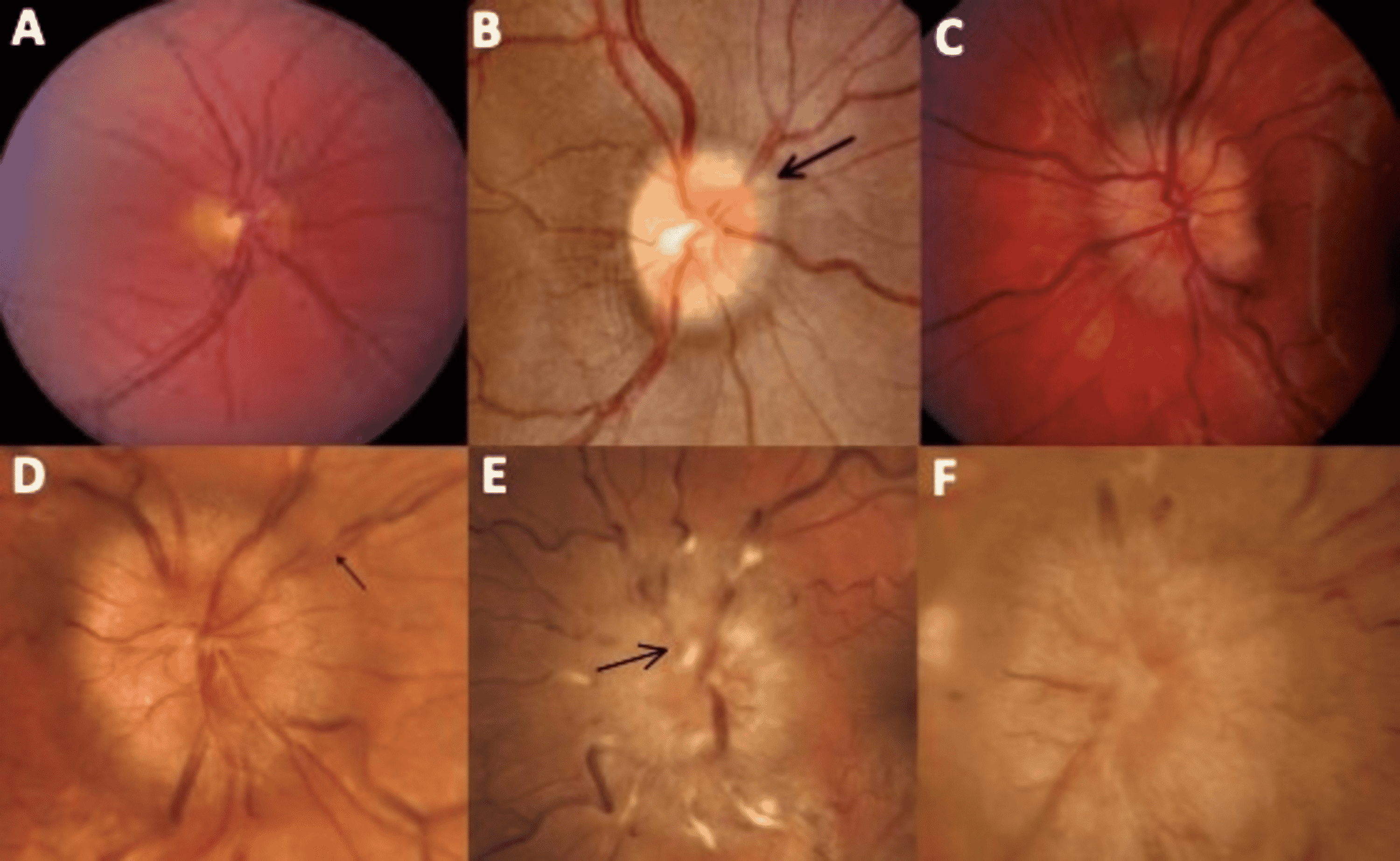
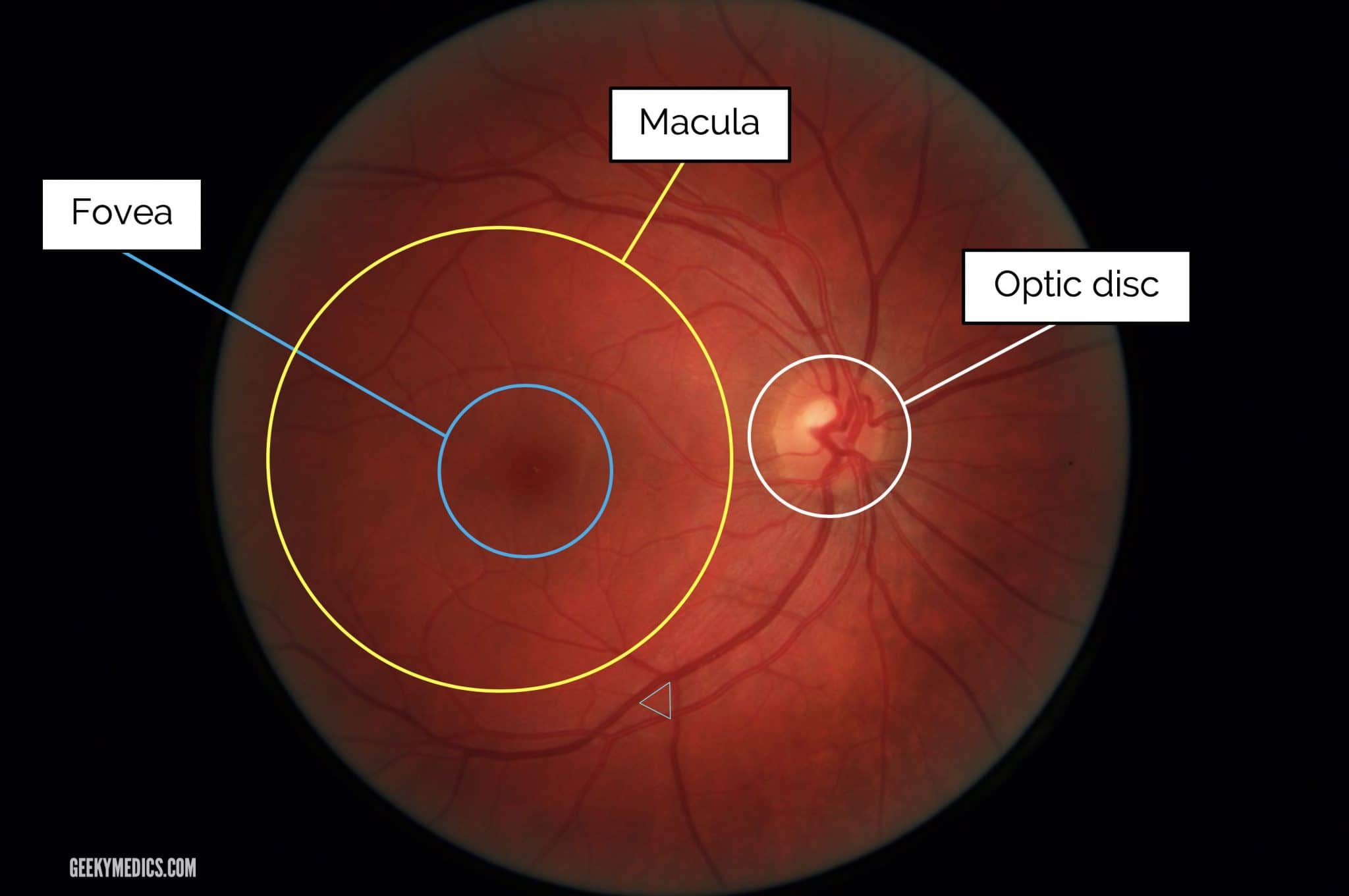
Diagnostic accuracy and use of nonmydriatic ocular fundus photography by emergency physicians: phase II of the FOTO-ED study. Am J Ophthalmol 2013 156 (5): 1056–1061 e10.īruce BB, Thulasi P, Fraser CL, Keadey MT, Ward A, Heilpern KL et al. Teaching ophthalmoscopy to medical students (the TOTeMS study). Kelly LP, Garza PS, Bruce BB, Graubart EB, Newman NJ, Biousse V. Ophthalmology in the medical school curriculum: reestablishing our value and effecting change. Undergraduate ophthalmology education - a survey of UK medical schools. Nonmydriatic ocular fundus photography in the emergency department. How effective is undergraduate and postgraduate teaching in ophthalmology? Eye 1997 11 (5): 744–750.īruce BB, Lamirel C, Wright DW, Ward A, Heilpern KL, Biousse V et al. The TOS study: can we use our patients to help improve clinical assessment? J R Coll Physicians Edinb 2012 42 (4): 306–310. Nicholl DJ, Yap CP, Cahill V, Appleton J, Willetts E, Sturman S. The second approach has clear advantages supported by evidence from the modern medical literature: it will empower non-ophthalmologists by allowing them to overcome that most significant hurdle of obtaining a useful fundal view and, ultimately and most importantly, improve patient safety and quality of care. There are two options to improve the quality of the examination: firstly commit more time and even more resources to teaching, and regularly assessing, direct ophthalmoscopy, which has been shown to be performed poorly even by experienced clinicians or, secondly, to routinely perform fundus photography which can be reviewed by the attending medical team and facilitate onward referral if required. 13 Simply attempting fundoscopy is not good enough-the examination needs to be reliable and accurate the documentation of an erroneous ‘normal’ fundus examination could certainly lead to harm. Considering such time restraints, rather than struggling to wield a direct ophthalmoscope in busy clinics, further clinical teaching is more of a priority for the modern medical student, which would enhance their ability to interpret signs from fundus images that can be captured by other means.įundus examination needs to be part of the complete physical examination as per the GMC and the Foundation Programme Curriculum. The time spent in ophthalmology by undergraduate medical students in the UK is limited, with most students spending 5–10 days of their entire training in compulsory ophthalmology education, 4 and trends across the developed world predict further reduction, 5 so it is vitally important that these precious hours are used wisely to ensure tomorrow’s doctors are best equipped to serve their patients. However, these signs must first be detected before they can be acted upon. The diagnosis of many systemic conditions can be made or assisted by the identification of ophthalmic features, and knowledge of ophthalmology in relation to acute medical and surgical problems in invaluable for any doctor. Ophthalmology-but not direct ophthalmoscopy-is an essential part of undergraduate medical education. 1, 2, 3 Missed or delayed detection of ophthalmic signs can result in harm to patients: undetected disc swelling could lead to loss of vision from persistently raised intracranial pressure an undiagnosed retinal artery occlusion could prevent potentially life-saving risk factor modification in a patient at risk of stroke failure to spot Roth spots could delay the diagnosis of bacterial endocarditis. Experienced physicians using the direct ophthalmoscope lack confidence and often fail to notice important abnormalities.


 0 kommentar(er)
